Bates’ Guide resources aid learning physical skills; test banks enhance comprehension through practice‚ mirroring real-world clinical scenarios for effective skill development․
Overview of the Test Bank
This test bank is meticulously designed to complement Bates’ Guide to Physical Examination‚ offering a robust collection of questions that assess understanding of core physical assessment techniques․ It features a diverse range of question types – multiple-choice‚ fill-in-the-blank‚ and case studies – to cater to varied learning styles and promote critical thinking․
The questions directly correlate with the content presented in the guide‚ reinforcing key concepts related to patient history taking‚ vital signs measurement‚ and systematic examination of body systems․
Instructors can utilize this resource to create customized quizzes and exams‚ while students benefit from self-assessment opportunities to identify areas needing further study and solidify their clinical skills․ The bank’s structure facilitates efficient learning and preparation for practical examinations․
Importance of Practice Questions
Consistent practice with these questions is paramount for mastering the intricacies of physical examination․ Repeated exposure to varied clinical scenarios strengthens recall and enhances the ability to apply theoretical knowledge to real patient encounters․ The test bank bridges the gap between textbook learning and practical application‚ fostering confidence in assessment skills․
Effective utilization of practice questions allows students to identify knowledge gaps and refine their diagnostic reasoning․
Furthermore‚ it simulates the exam environment‚ reducing test anxiety and improving performance․ Regular self-testing reinforces learning‚ solidifying understanding of key concepts and promoting long-term retention of vital clinical skills․
Target Audience: Students and Practitioners
This Bates’ Guide test bank is meticulously designed for both medical students honing their foundational skills and seasoned practitioners seeking continuing education or skill refinement․ Students benefit from the comprehensive coverage of examination techniques‚ while practitioners utilize it for staying current with best practices and reinforcing core competencies․
The resource caters to diverse learning styles‚ offering a flexible approach to mastering physical assessment․
It’s invaluable for residency programs‚ clinical rotations‚ and board preparation‚ ensuring a strong clinical foundation for all healthcare professionals․
Cardiovascular System Examination
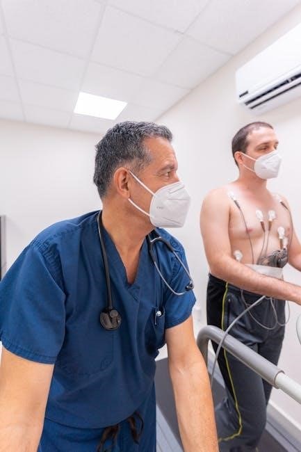
Bates’ Guide focuses on vital signs‚ auscultation‚ and pulse palpation to assess cardiac function‚ identifying abnormalities and guiding diagnostic evaluations effectively․
Vital Signs Assessment
Bates’ Guide emphasizes a systematic approach to vital signs‚ beginning with accurate temperature measurement – oral‚ tympanic‚ axillary‚ or rectal – noting any deviations from normal ranges․ Pulse rate assessment involves palpation of radial or carotid arteries‚ evaluating rate‚ rhythm‚ and amplitude․
Respiration rate is observed‚ noting depth and regularity‚ while blood pressure measurement utilizes auscultation with a stethoscope‚ documenting systolic and diastolic pressures․ Bates’ stresses the importance of considering patient factors like age‚ stress‚ and medications when interpreting vital signs․
Accurate documentation and recognizing trends are crucial for identifying potential cardiovascular or respiratory compromise‚ guiding further examination and intervention․ Mastering these foundational skills is paramount for effective patient care․
Auscultation of Heart Sounds
Bates’ Guide details a precise technique for auscultating heart sounds‚ utilizing the diaphragm of the stethoscope at specific auscultatory areas: aortic‚ pulmonic‚ tricuspid‚ and mitral․ Identifying S1‚ the closure of the mitral and tricuspid valves‚ and S2‚ the closure of the aortic and pulmonic valves‚ forms the basis of normal heart sounds․
Bates’ thoroughly explains recognizing abnormal sounds like murmurs – categorized by timing‚ pitch‚ and intensity – and gallops (S3 and S4)‚ indicative of ventricular dysfunction․
Distinguishing between innocent murmurs and pathological ones requires careful assessment and correlation with clinical findings․ Proper technique and a systematic approach‚ as outlined in Bates’‚ are essential for accurate interpretation․
Normal Heart Sounds (S1 and S2)
Bates’ Guide emphasizes S1‚ a low-pitched sound coinciding with the start of ventricular systole‚ caused by mitral and tricuspid valve closure․ Its intensity varies with factors like heart rate and position․ S2‚ a higher-pitched sound‚ marks the beginning of diastole‚ resulting from aortic and pulmonic valve closure․
Bates’ clarifies that S2 normally splits during inspiration due to increased venous return and delayed pulmonic valve closure․ The timing and character of this split are clinically significant․
Accurate identification of S1 and S2‚ as detailed in Bates’‚ is fundamental to differentiating normal from abnormal heart sounds and forming a basis for further cardiac assessment․
Abnormal Heart Sounds (Murmurs‚ Gallops)
Bates’ Guide details murmurs as prolonged extra sounds‚ categorized by timing (systolic‚ diastolic) and intensity (graded I-VI)․ Systolic murmurs often indicate valve stenosis or regurgitation․ Diastolic murmurs frequently suggest valve problems like mitral stenosis․
Bates’ explains gallops – S3 and S4 – as extra sounds indicating ventricular dysfunction․ S3‚ a low-pitched sound early in diastole‚ suggests volume overload․ S4‚ a higher-pitched sound before S1‚ indicates decreased ventricular compliance․
Properly identifying these abnormal sounds‚ as taught by Bates’‚ requires careful auscultation and differentiation from innocent or functional murmurs‚ guiding further diagnostic evaluation․
Palpation of Peripheral Pulses
Bates’ Guide emphasizes peripheral pulse palpation as crucial for assessing arterial blood flow and cardiovascular health․ The technique involves gently pressing on arteries to feel pulse strength – graded 0 to 4+․
Bates’ details key pulse locations: radial (wrist)‚ dorsalis pedis (foot)‚ posterior tibial (ankle)‚ brachial (arm)‚ and femoral (groin)․ Assessing symmetry‚ rate‚ and rhythm is vital․ Diminished or absent pulses suggest arterial obstruction․
Bates’ instructs clinicians to correlate pulse findings with patient history and other examination results․ Accurate pulse assessment‚ as outlined in Bates’‚ aids in diagnosing peripheral artery disease and other vascular conditions․
Radial Pulse Assessment
Bates’ Guide details radial pulse assessment‚ a foundational skill․ Locate the pulse on the thumb side of the wrist․ Use the pads of your index and middle fingers‚ avoiding the thumb’s pulse․
Bates’ instructs to assess rate (beats per minute)‚ rhythm (regularity)‚ and amplitude (strength – graded 0 to 4+)․ A normal rate is 60-100 bpm․ Irregular rhythms warrant further investigation․
Bates’ emphasizes comparing both radial pulses for symmetry․ Weakness or absence suggests potential vascular issues․ Document findings precisely‚ as per Bates’ guidelines‚ for accurate patient evaluation and informed clinical decision-making․
Dorsalis Pedis and Posterior Tibial Pulse Assessment
Bates’ Guide outlines assessing dorsalis pedis (top of foot) and posterior tibial (behind medial malleolus) pulses․ Palpate with index and middle fingers‚ noting rate‚ rhythm‚ and amplitude (0-4+ scale)․
Bates’ stresses comparing pulses bilaterally․ Diminished or absent pulses indicate potential peripheral artery disease․ The dorsalis pedis can be difficult to locate in some patients; Bates’ suggests techniques for enhancement․
Bates’ emphasizes accurate documentation․ Pulse assessment‚ as detailed by Bates’‚ is crucial for evaluating vascular sufficiency․ Consider patient factors like age and comorbidities when interpreting findings‚ following Bates’ comprehensive approach․
Respiratory System Examination
Bates’ Guide details inspecting‚ palpating‚ percussing‚ and auscultating the lungs‚ identifying normal and abnormal breath sounds for comprehensive respiratory assessment․
Inspection of Respiratory Effort
Bates’ Guide emphasizes a systematic approach to observing respiratory patterns․ Begin by noting the respiratory rate‚ rhythm‚ and depth․ Observe for signs of labored breathing‚ such as nasal flaring‚ retractions (intercostal‚ supraclavicular‚ or suprasternal)‚ and the use of accessory muscles – sternocleidomastoid and scalene muscles․
Assess the patient’s position; are they orthopneic (preferring to sit upright) or tripodding (leaning forward with hands on knees)? Look for cyanosis‚ indicating inadequate oxygenation․ Chest expansion should be symmetrical; asymmetry may suggest underlying pathology like pneumothorax or pleural effusion․ Document any visible deformities of the chest wall․ A thorough inspection provides crucial initial clues about respiratory function before proceeding to further examination techniques․
Auscultation of Lung Sounds
Bates’ Guide details a precise technique for auscultating lung sounds․ Use the diaphragm of the stethoscope‚ instructing the patient to breathe deeply through their mouth․ Systematically compare sounds in all lung fields‚ anterior‚ posterior‚ and lateral‚ noting any differences․
Normal breath sounds include vesicular sounds (soft‚ breezy) heard over most lung fields‚ and bronchial sounds (louder‚ harsher) over the trachea․ Identify any adventitious (abnormal) sounds like wheezes (high-pitched whistling)‚ crackles (discontinuous popping)‚ or rhonchi (low-pitched snoring)․ Document the timing‚ location‚ and characteristics of any abnormal sounds encountered during auscultation․
Normal Breath Sounds (Vesicular‚ Bronchial)
Bates’ Guide emphasizes differentiating normal breath sounds․ Vesicular sounds are soft‚ low-pitched‚ and heard over most lung fields‚ representing quiet airflow in the alveoli․ They are longer on inspiration than expiration․ Bronchial sounds‚ conversely‚ are louder‚ higher-pitched‚ and have an equal duration of inspiration and expiration․
These are normally heard over the trachea and main bronchi․ Recognizing these distinctions is crucial․ Deviations from these expected sounds—increased or decreased intensity‚ altered timing—indicate potential respiratory issues․ Accurate identification requires careful listening and systematic comparison across lung fields‚ as detailed within the guide․
Adventitious Breath Sounds (Wheezes‚ Crackles)
Bates’ Guide thoroughly covers adventitious‚ or abnormal‚ breath sounds․ Wheezes are continuous‚ high-pitched‚ whistling sounds‚ often indicative of airway narrowing‚ like in asthma or COPD․ Crackles‚ formerly called rales‚ are discontinuous‚ popping sounds heard during inspiration‚ suggesting fluid in the small airways‚ potentially from pneumonia or heart failure․
The guide stresses differentiating these sounds by timing‚ pitch‚ and location․ Recognizing these abnormal sounds requires focused auscultation and correlating findings with the patient’s clinical presentation․ Proper identification aids in accurate diagnosis and guides appropriate treatment strategies‚ as emphasized by Bates’ methodology․
Percussion of Lung Fields
Bates’ Guide details percussion as a vital technique for assessing underlying lung tissue․ Percussion involves tapping the chest wall to evaluate resonance – the hollow sound of normal lung tissue․ Dullness suggests increased density‚ like in pneumonia or pleural effusion‚ while hyperresonance indicates air trapping‚ as seen in emphysema․
The guide emphasizes systematic percussion‚ comparing sounds bilaterally to identify abnormalities․ Skilled percussion‚ combined with auscultation‚ provides crucial information about lung density and potential pathology․ Bates’ stresses that percussion isn’t just about identifying what’s wrong‚ but confirming what’s normal as a baseline․

Neurological Examination
Bates’ Guide emphasizes a systematic neurological assessment‚ covering mental status‚ cranial nerves‚ motor/sensory functions‚ and reflexes for comprehensive evaluation․
Mental Status Assessment
Bates’ Guide details a thorough mental status examination‚ crucial for identifying cognitive and emotional alterations․ This assessment begins with observing the patient’s appearance and behavior‚ noting any obvious distress or unusual mannerisms․
Next‚ evaluate the patient’s level of consciousness – are they alert‚ lethargic‚ or comatose? Assess orientation to person‚ place‚ and time․ Then‚ examine speech and language‚ looking for fluency‚ comprehension‚ and any aphasias․ Memory assessment‚ including recent and remote recall‚ is vital․
Finally‚ evaluate thought process‚ mood‚ and affect․ Look for logical thought patterns and appropriate emotional responses․ Document any inconsistencies or abnormalities observed during this comprehensive evaluation‚ as these findings can provide critical clues to underlying neurological or psychiatric conditions․
Cranial Nerve Examination
Bates’ Guide systematically outlines the cranial nerve examination‚ essential for pinpointing neurological dysfunction․ Testing begins with Olfactory (I) – identifying familiar scents with each nostril occluded․ Optic Nerve (II) assessment involves visual acuity‚ visual fields‚ and fundoscopic examination to evaluate the optic disc and retina․
Oculomotor (III)‚ Trochlear (IV)‚ and Abducens (VI) nerves are tested by assessing extraocular movements in all directions of gaze․ Facial (VII) nerve function is evaluated through facial expressions and taste sensation․ Acoustic (VIII) nerve testing assesses hearing and balance․
Glossopharyngeal (IX) and Vagus (X) nerves are examined via gag reflex and palate elevation․ Accessory (XI) nerve strength is tested by shoulder shrug and head rotation‚ while Hypoglossal (XII) nerve function is assessed by tongue protrusion․
Testing of Olfactory Nerve (I)
Bates’ Guide details olfactory nerve (I) testing as a crucial initial step․ Each nostril is assessed separately‚ occluding the other to prevent carryover․ Use non-irritating substances – coffee grounds‚ vanilla‚ or lemon peel are preferred‚ avoiding strong scents like ammonia․
The patient closes their eyes and identifies the scent held briefly under each nostril․ Anosmia (loss of smell) or hyposmia (reduced smell) indicates potential dysfunction․ Document which nostril‚ if any‚ demonstrates impairment․
False positives can occur with nasal congestion or allergies․ Always inquire about recent upper respiratory infections or nasal surgery‚ as these can affect results․ Safety is paramount; avoid testing if there’s a risk of aspiration․
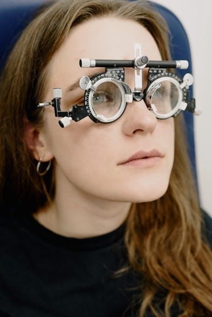
Testing of Optic Nerve (II)
Bates’ Guide emphasizes a systematic approach to optic nerve (II) assessment․ Begin with visual acuity‚ using a Snellen chart at 20 feet‚ testing each eye individually and then both together․ Note any corrective lenses used․
Next‚ assess visual fields via confrontation․ The examiner focuses on a central point while the patient fixates‚ and peripheral vision is tested in each quadrant․ Assess pupillary reactions to light – direct and consensual – observing for size‚ shape‚ and symmetry․
Fundoscopic examination reveals the optic disc‚ retina‚ and vessels․ Look for pallor‚ edema‚ or hemorrhages․ Document any abnormalities meticulously‚ as they can indicate various neurological conditions․
Motor and Sensory Examination
Bates’ Guide details a thorough motor and sensory assessment․ Begin with observation for spontaneous movements‚ symmetry‚ and any involuntary motions․ Test muscle strength using a standardized scale (0-5) across major muscle groups‚ comparing left to right․ Assess tone by gently flexing and extending limbs‚ noting resistance․
Sensory testing involves light touch‚ pain‚ temperature‚ and vibration․ Utilize a symmetrical approach‚ comparing corresponding areas on both sides of the body․ Assess proprioception (joint position sense) and graphesthesia (letter recognition)․
Document any deficits precisely‚ noting location and type‚ to aid in neurological localization․
Muscle Strength Testing
Bates’ Guide emphasizes a systematic approach to muscle strength testing․ Grade strength on a 0-5 scale: 0 (no contraction)‚ 1 (flicker)‚ 2 (movement with gravity eliminated)‚ 3 (movement against gravity)‚ 4 (good strength)‚ and 5 (normal strength)․
Test key muscle groups bilaterally‚ comparing sides․ Stabilize the proximal segment and ask the patient to resist your force․ Observe for fatigue or pain․ Document the grade for each muscle group tested․
Consider factors like patient effort and cooperation․ Strength deficits can indicate neuromuscular disorders or nerve damage‚ guiding further investigation․
Assessment of Reflexes
Bates’ Guide details a standardized reflex assessment․ Utilize a reflex hammer to elicit deep tendon reflexes (DTRs) – biceps‚ triceps‚ brachioradialis‚ patellar‚ and Achilles․ Grade reflexes on a scale of 0 (absent)‚ 1+ (hypoactive)‚ 2+ (normal)‚ 3+ (brisk)‚ and 4+ (hyperactive)․
Ensure the patient is relaxed․ Compare responses bilaterally․ Asymmetry or abnormal grading warrants further investigation․ Reinforcement maneuvers can enhance responses if initially diminished․
Assess for pathological reflexes like Babinski’s sign (upgoing plantar response)‚ indicating upper motor neuron damage․ Document findings clearly‚ noting any abnormalities for accurate neurological evaluation․
Abdominal Examination
Bates’ Guide emphasizes systematic abdominal assessment: inspection‚ auscultation‚ percussion‚ and palpation‚ identifying tenderness‚ masses‚ and organomegaly effectively․
Inspection of Abdomen
Bates’ Guide details a thorough abdominal inspection‚ beginning with skin assessment for scars‚ striae‚ and vascular patterns․ Observe for symmetry‚ contour – flat‚ rounded‚ scaphoid – and any visible pulsations or peristalsis․
Note any distension‚ masses‚ or hernias․ Assess for visible respiratory movement‚ noting if it’s symmetrical․ The examiner should evaluate for signs of referred pain or inflammation․ Proper lighting is crucial for accurate observation․
Patient positioning – typically supine with knees bent – facilitates relaxation of abdominal muscles‚ enhancing the inspection process․ This initial visual assessment provides valuable clues before proceeding to auscultation‚ percussion‚ and palpation‚ forming a foundational step in the abdominal examination․
Auscultation of Bowel Sounds
Bates’ Guide emphasizes auscultation before percussion and palpation‚ as these maneuvers can alter bowel activity․ Use the diaphragm of the stethoscope‚ listening in all four quadrants․ Normal bowel sounds are irregular‚ gurgling‚ and varying in pitch and loudness․
Document the character and frequency – absent‚ hypoactive‚ normoactive‚ or hyperactive․ Borborygmi‚ loud prolonged gurgles‚ indicate increased motility․ Absent sounds require further investigation․
Listen for at least five minutes in each quadrant before concluding sounds are absent․ Consider vascular bruits‚ which suggest arterial stenosis․ Accurate documentation of bowel sounds is crucial for diagnosing various gastrointestinal conditions and monitoring patient response to treatment․
Palpation of Abdomen
Bates’ Guide details a systematic approach to abdominal palpation‚ beginning with light palpation to assess for tenderness‚ muscle guarding‚ and superficial organomegaly․ Use circular motions‚ progressing across all quadrants․ Then‚ perform deep palpation to detect deeper structures and masses‚ ensuring the patient is relaxed․
Note any areas of resistance or pain․ Assess for rebound tenderness‚ indicating peritoneal inflammation․ Palpate for the liver‚ spleen‚ and kidneys‚ documenting size‚ shape‚ and tenderness․
Proper technique minimizes patient discomfort and maximizes diagnostic yield‚ aiding in the identification of abdominal pathologies․
Light and Deep Palpation

Bates’ Guide emphasizes differentiating light and deep palpation․ Light palpation‚ using gentle circular motions with the pads of the fingers‚ assesses for superficial tenderness‚ muscle guarding‚ and surface abnormalities․ It’s the initial step‚ establishing a baseline․
Deep palpation employs firmer pressure‚ using the palmar surface of the hands‚ to detect deeper organs‚ masses‚ or areas of resistance․ The examiner should press inward and downward‚ feeling for the liver‚ spleen‚ and kidneys․
Comparing findings between quadrants is crucial for identifying localized pathology․ Patient comfort is paramount throughout both techniques․
Assessment for Organomegaly
Bates’ Guide details a systematic approach to detecting organomegaly․ Palpation focuses on the liver‚ spleen‚ and kidneys‚ noting size‚ shape‚ and tenderness․ Liver size is assessed by measuring the distance from the right costal margin‚ accounting for respiratory movement․
Splenic enlargement is often felt in the left upper quadrant‚ potentially indicating infection or hematologic disorders․ Kidney assessment involves bimanual palpation‚ attempting to trap the kidney between the hands․
Accurate documentation of any palpable enlargement‚ along with associated findings‚ is essential for diagnosis and management․

Musculoskeletal System Examination
Bates’ Guide emphasizes evaluating joint structure‚ range of motion‚ and muscle strength‚ identifying pain‚ swelling‚ and deformity for accurate musculoskeletal assessment․
Range of Motion Assessment
Bates’ Guide details a systematic approach to assessing range of motion (ROM) in all major joints․ This involves observing active ROM – the patient’s ability to move the joint independently – and passive ROM‚ where the examiner moves the joint through its arc․
The guide stresses noting any limitations‚ pain‚ or crepitus during movement․ ROM is typically assessed using goniometry‚ a precise measurement technique․ Understanding normal ROM values is crucial for identifying abnormalities․
Assessments should include flexion‚ extension‚ abduction‚ adduction‚ internal and external rotation‚ where applicable․ Documenting findings accurately is paramount for tracking progress and informing treatment plans․ A comprehensive ROM assessment is fundamental to a thorough musculoskeletal examination․
Joint Examination
Bates’ Guide emphasizes a four-step approach to joint examination: inspection‚ palpation‚ range of motion testing‚ and special tests․ Inspection focuses on observing for swelling‚ redness‚ deformity‚ or muscle atrophy․ Palpation assesses for tenderness‚ warmth‚ and effusion within the joint capsule․
Following ROM assessment‚ specific maneuvers are performed to evaluate ligamentous stability and identify potential pathology․ These special tests often provoke symptoms or demonstrate abnormal movement․
Accurate documentation of findings‚ including location and character of pain‚ is vital․ A systematic joint examination‚ guided by Bates’ principles‚ allows for precise diagnosis and effective management of musculoskeletal conditions․
Inspection for Swelling and Deformity
Bates’ Guide stresses careful visual assessment during joint inspection․ Look for any noticeable swelling‚ which can indicate effusion or inflammation within the joint․ Compare the examined joint to the contralateral side for symmetry․ Deformities‚ such as angulation or subluxation‚ should be meticulously noted‚ documenting their location and degree․
Observe the skin overlying the joint for redness‚ bruising‚ or any signs of trauma․ Muscle atrophy can also be visible‚ suggesting chronic disuse or nerve involvement․
A systematic inspection‚ as outlined in Bates’‚ provides crucial initial clues to underlying joint pathology‚ guiding further examination steps;
Palpation for Tenderness
Bates’ Guide emphasizes a systematic approach to palpation‚ beginning with light touch to assess superficial tissues․ Progress to deeper palpation‚ using the pads of your fingers to detect areas of tenderness․ Note any localized pain‚ crepitus‚ or muscle spasm․ Palpate bony landmarks‚ ligaments‚ and tendons surrounding the joint․
Ask the patient to report any discomfort during palpation‚ characterizing the pain’s location‚ intensity‚ and quality․ Compare findings to the contralateral joint to identify asymmetries․
Careful palpation‚ as detailed in Bates’‚ helps pinpoint the source of musculoskeletal pain and guides further diagnostic evaluation․